VN
LÝ THUYẾT VÀ ÁP DỤNG PHƯƠNG PHÁP DỰ ĐOÁN TĂNG TRƯỞNG THEO RICKETTS
1.Tổng quan và các vị trí quyết định tăng trưởng theo Ricketts
Robert Murray Ricketts (1920 – 2003) là giáo sư chỉnh nha được biết đến với những đóng góp ý nghĩa cho ngành nắn chỉnh răng như phát triển phân tích phim sọ nghiêng Ricketts, phát triển triết lý Bioprogressive, hệ thống mắc cài slot .018 inch, cung tiện ích Ricketts,…
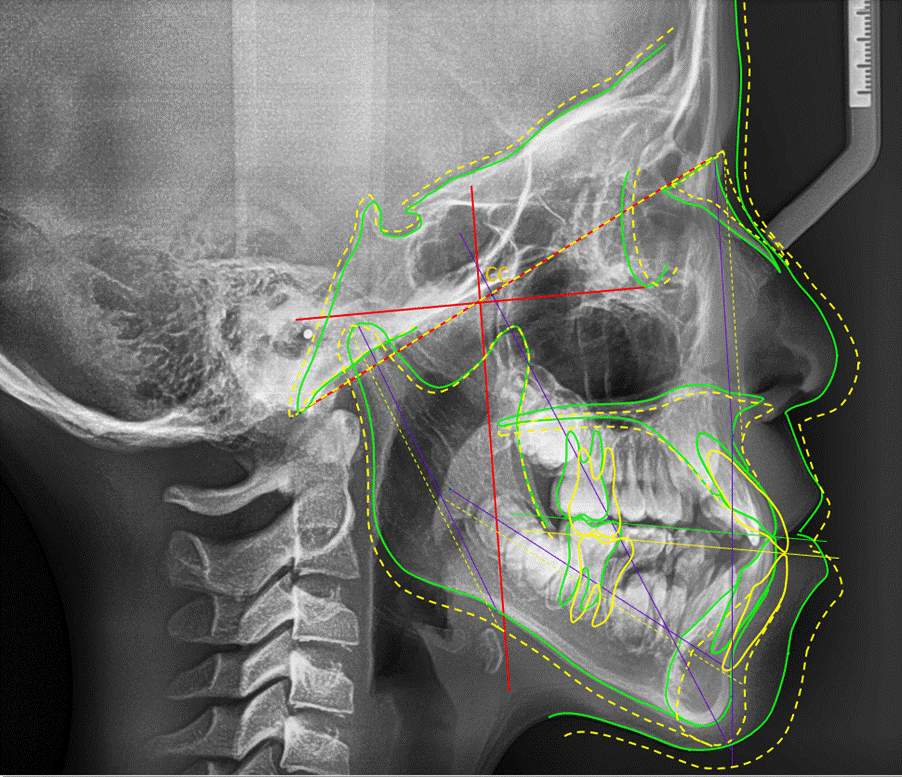
Phân tích phim sọ nghiêng của Ricketts chú trọng đến yếu tố tăng trưởng, giá trị chuẩn của các chỉ số được Ricketts công bố theo giới tính, chủng tộc và độ tuổi. Năm 1957, Ricketts đưa ra quy trình dự đoán tăng trưởng. Ba yếu tố chính tạo nên những khác biệt về hình thái tăng trưởng đã được Ricketts đề cập đến bao gồm 1
- Thay đổi ở nền sọ
- Thay đổi vị trí lồi cầu
- Sự phát triển ở lồi cầu về cả lượng và hướng
2.Các bước thực hành dự đoán tăng trưởng
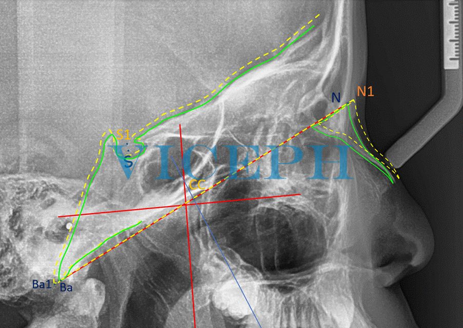
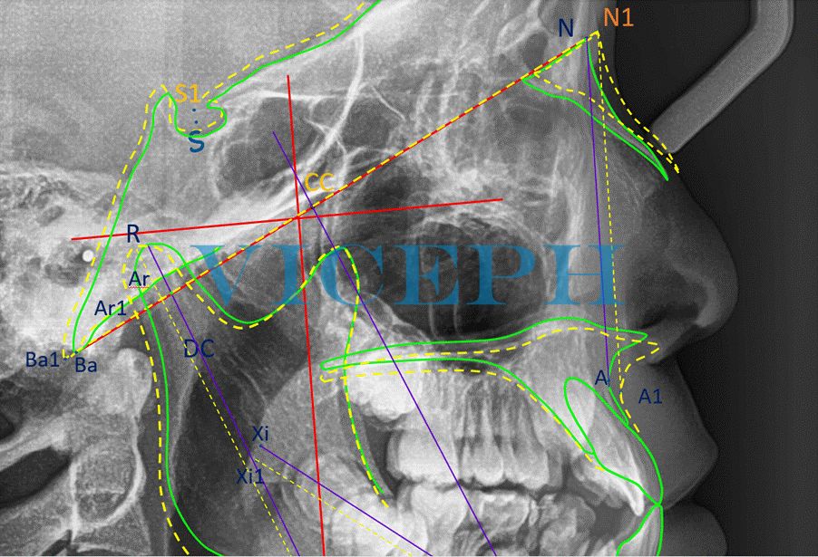
2.1. Thay đổi tại nền sọ
Yếu tố đầu tiên được xem xét tới là sự thay đổi ở nền sọ, được cho là ít quan trọng nhất trong quá trình điều trị.
Theo nghiên cứu của Ricketts, hầu như giá trị góc NSBa không thay đổi nhưng nền sọ tăng trưởng cả về phía trước và phía sau trong thời kỳ tăng trưởng, cụ thể SN tăng 1mm mỗi năm, SBa tăng 0.75mm mỗi năm. 1, 2
Để đơn giản hơn, người ta lấy Ba-N (mặt phẳng nền sọ) như một trục phát triển, và theo trục này, N ra trước 1mm mỗi năm và Ba lùi sau 1mm mỗi năm. 3
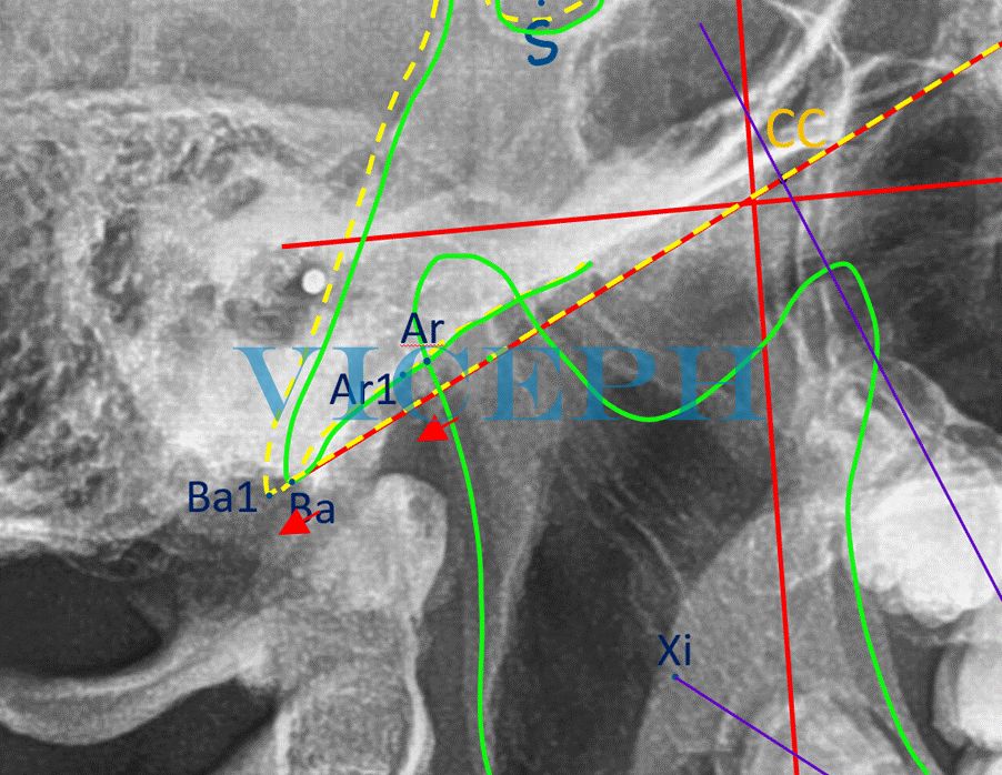
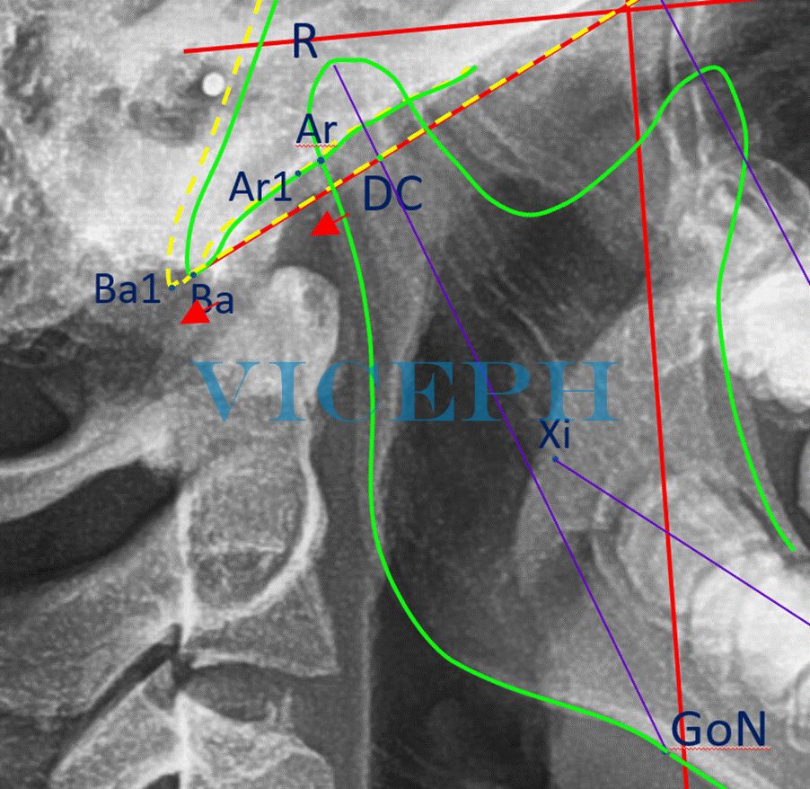
2.2. Thay đổi vị trí của lồi cầu
Yếu tố thứ hai ảnh hưởng đến hình thái tăng trưởng của khuôn mặt là sự thay đổi vị trí của lồi cầu.
Khoảng 60% trường hợp được theo dõi cho thấy vị trí lồi cầu không thay đổi. Hơn thế nữa, trong suốt quá trình phát triển, điểm Ar (giao điểm của Ba-N với bờ sau lồi cầu) chuyển động xuống dưới và ra sau song song với chuyển động của Ba.1, 2
Từ dữ liệu trên, chúng ta có thể sẽ xác định được vị trí dịch chuyển của lồi cầu theo điểm Ba.
2.3. Thay đổi phát triển lồi cầu
Yếu tố thứ 3 và là yếu tố quan trọng nhất đối với sự phát triển hình thái khuôn mặt là sự phát triển của lồi cầu.
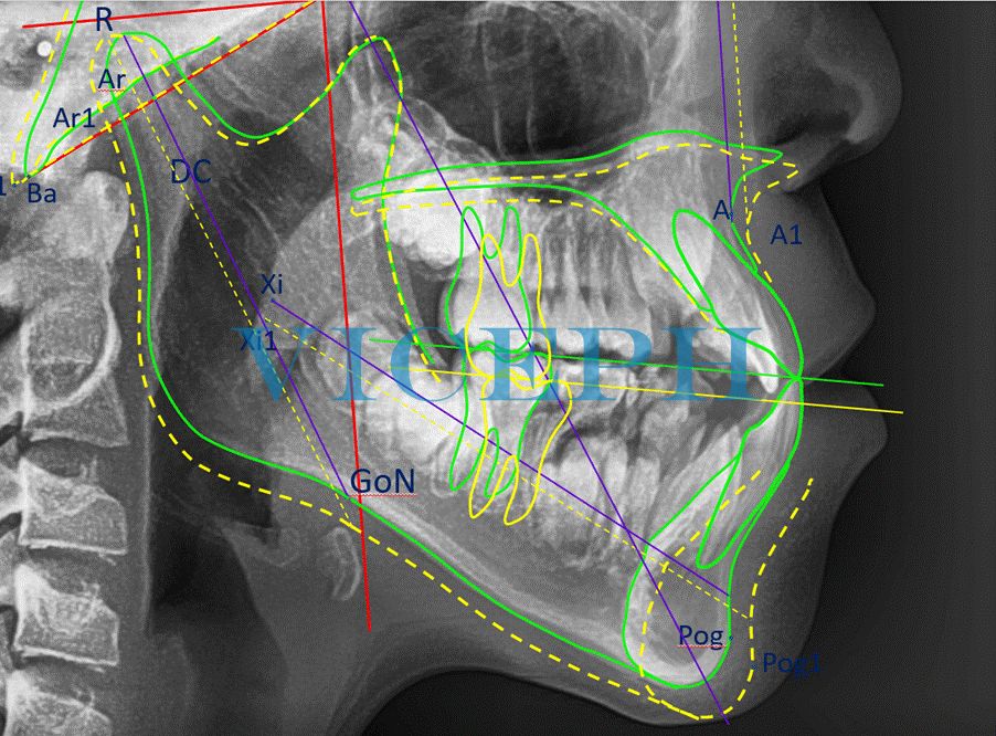
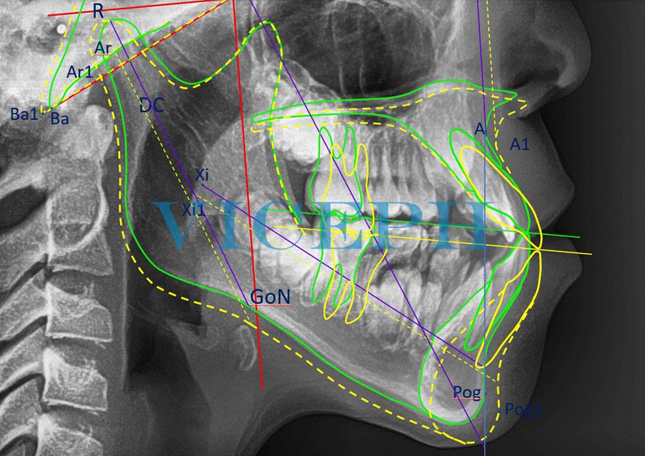
Để dự đoán tăng trưởng của lồi cầu, Ricketts đưa ra khái niệm trục lồi cầu. Trục lồi cầu được định nghĩa là trục dài đi qua tâm lồi cầu (DC) và điểm sâu nhất của lõm trước góc hàm (GoN).1
Các thay đổi cụ thể bao gồm:
- Về góc độ, trục lồi cầu thường thay đổi trong quá trình điều trị, thường là xoay mở gây cắn hở quá mức với trường hợp góc hàm dưới lớn, góc hàm kém nhô, chỏm lồi cầu ngắn, cành cao mỏng; xoay đóng trong trường hợp góc Gonial nhọn, thân hàm dưới phát triển tốt, cành cao dày, chỏm lồi cầu phát triển. Tuy nhiên, trong quá trình phát triển không có can thiệp điều trị, trục này thường ít thay đổi, góc giữa trục lồi cầu và mặt phẳng Frankfort chỉ giảm khoảng 0.3 độ/năm.
- Về độ dài, độ dài của trục lồi cầu tăng khoảng 2mm/năm.1, 2
Với sự thay đổi về góc độ và kích thước của trục lồi cầu, ta có thể dự đoán hình thái phát triển của cành lên, kết thúc tại GoN. Điểm GoN là điểm nối giữa cành lên và thân xương hàm dưới, là khu vực tuyệt vời để điều chỉnh trong dự đoán tăng trưởng hàm dưới. 1
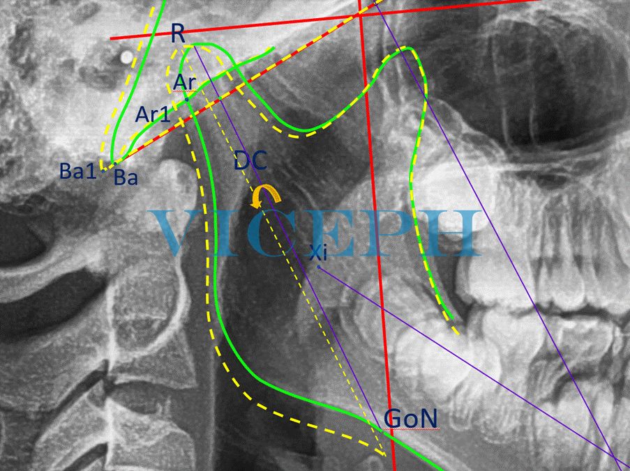
Tăng trưởng của thân xương hàm dưới tiếp tục được dự đoán dựa vào độ dài thân XHD (Xi-Pm) tăng 1,6mm/ năm và góc giữ Xi-Pm và DC-Xi đóng 0.5 độ mỗi năm. 4
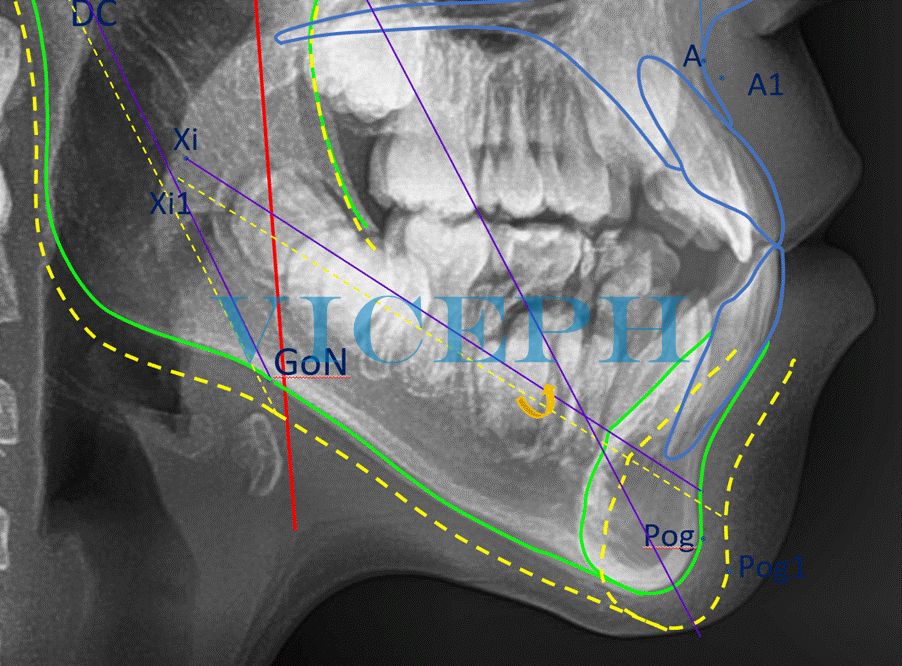
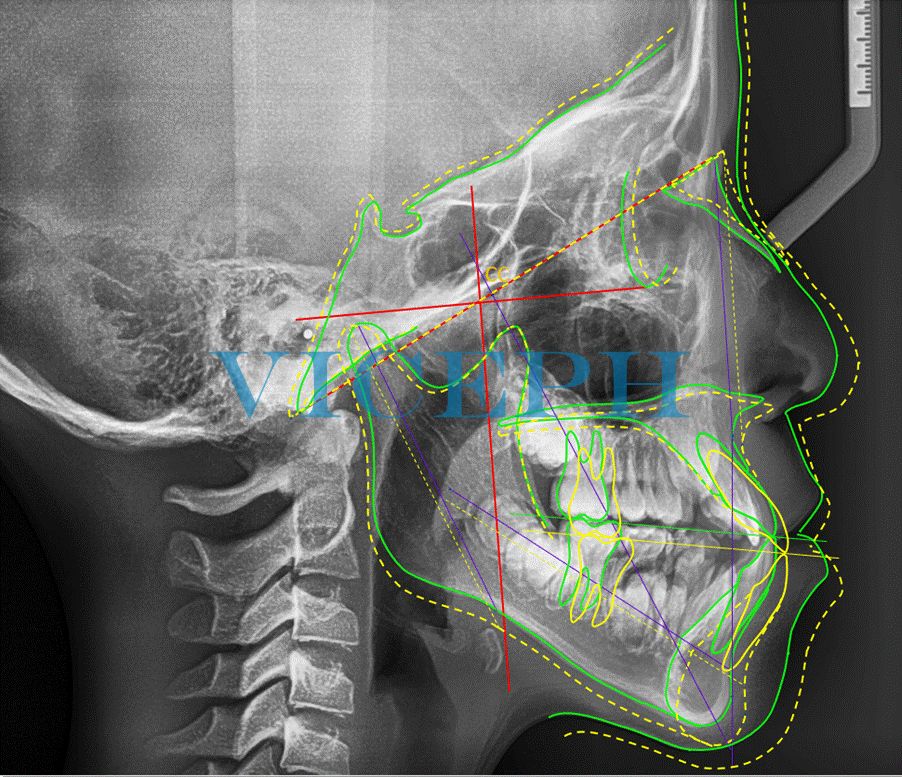
Sau khi hoàn thiện dự đoán tăng trưởng của 3 yếu tố chính ảnh hưởng đến sự khác biệt của hình thái khuôn mặt, cần hoàn thiện các phần còn lại: hàm trên, răng và mô mềm.
Ricketts chỉ ra rằng, độ tăng của tổng chiều cao mặt 40% là do sự tăng chiều cao của tầng mặt giữa và 60% là do sự tăng chiều cao của tầng mặt dưới.1 Vì vậy, vị trí mới của điểm A và XHT được xác định dựa trên góc SNA không thay đổi 1, 2 và NA tăng bằng 40% độ tăng tổng chiều cao mặt.
2.4. Các dịch chuyển của răng
Với răng hàm lớn, chúng sẽ dịch chuyển theo cả chiều ngang và chiều dọc: chiều ngang dịch chuyển ra trước 1mm/năm;4 chiều dọc dịch chuyển theo XHT và thêm ½ độ tăng chiều cao tầng mặt dưới,1 tức là chiều dọc dịch chuyển bằng 70% độ tăng tổng chiều cao mặt.
Với các răng cửa, đầu tiên dịch chuyển chúng giống răng hàm lớn, tiếp đó xoay trục các răng cửa sao cho góc giữa trục răng cửa và APog không thay đổi so với ban đầu.
2.5. Dịch chuyển mô mềm
Ricketts không đề cập nhiều đến tăng trưởng của mô mềm mà chủ yếu đưa ra tăng trưởng của mũi. Đỉnh mũi (Prn) tăng trưởng về phía trước so với ANS 1mm mỗi năm.1, 2
Hoàn thiện phần tăng trưởng của mũi, ta có được kết quả dự đoán tăng trưởng.
3. Kết luận
Kết quả tăng trưởng không phải luôn luôn trùng khớp với tăng trưởng thật sự nhưng có nhiều giá trị tham khảo.
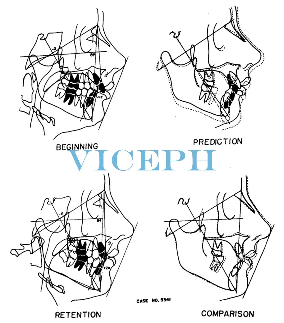
Việc dự đoán tăng trưởng khi kết hợp với việc lập kế hoạch điều trị (VTO-Visual Treatment Objective) cho phép bác sĩ chỉnh nha dự đoán kết quả sau điều trị trong quá trình tăng trưởng.
Dưới đây là hình ảnh VTO dựa trên dự đoán tăng trưởng được Ricketts công bố.
EN
Theory and Application of Ricketts’ Growth Prediction Method
1. Overview and Determining Factors of Growth According to Ricketts
Robert Murray Ricketts (1920 – 2003) was a renowned orthodontist known for his significant contributions to the field of orthodontics, such as the development of Ricketts’ cephalometric analysis, the Bioprogressive philosophy, the .018-inch slot bracket system, and Ricketts’ utility arch.
Ricketts’ cephalometric analysis focused on growth factors, and he published standardized values of various indices based on gender, ethnicity, and age. In 1957, Ricketts introduced a growth prediction procedure. He identified three main factors that contribute to differences in growth morphology, which include:1
- Changes in the cranial base
- Changes in convexity position
- Development of convexity in terms of both magnitude and direction
2. Practice Steps for Growth Prediction
2.1. Changes in the Cranial Base
The first factor considered is the changes in the cranial base, which are believed to be the least important in the treatment process.
According to Ricketts’ research, the NSBa angle remains almost constant, but the cranial base undergoes growth both anteriorly and posteriorly during the growth period. Specifically, SN increases by 1mm per year, and SBa increases by 0.75mm per year. 1,2
To simplify, Ba-N (cranial base plane) is taken as a developmental axis, and along this axis, N moves forward by 1mm per year, while Ba moves backward by 1mm per year. 3
2.2. Changes in the Position of Convexity
The second factor influencing the growth morphology of the face is the changes in the position of convexity.
Approximately 60% of the observed cases show no change in the position of convexity. Moreover, during the development process, the point Ar (intersection of Ba-N with the posterior border of convexity) moves downward and posteriorly parallel to the movement of Ba. 1,2
Based on the above data, we can determine the shifting position of convexity using the Ba point.
2.3. Developmental Changes in Convexity
The third and most significant factor for the development of facial morphology is the development of convexity.
To predict the growth of convexity, Ricketts introduced the concept of the convexity axis. The convexity axis is defined as a long axis passing through the center of convexity (DC) and the deepest point of the concavity in the anterior angle of the jaw (GoN).1
The specific changes include:
- In terms of angle, the convexity axis typically undergoes changes during the treatment process. It often rotates open, causing excessive overbite in cases of large lower jaw angle, deficient jaw projection, short convexity chamfer, and high, thin ramus. Conversely, it rotates closed in cases of sharp Gonial angle, well-developed lower jaw body, thick ramus, and developed convexity chamfer. However, without treatment intervention, this axis tends to change minimally, with the angle between the convexity axis and the Frankfort plane decreasing by approximately 0.3 degrees per year.
- In terms of length, the length of the convexity axis increases by approximately 2mm per year. 1,2
With changes in the angle and size of the convexity axis, we can predict the developmental morphology of the ascending branch, ending at GoN. The GoN point is the junction point between the ascending branch and the lower jaw body, and it is an excellent area for adjustment in growth prediction of the lower jaw. 1
The growth of the lower jaw body continues to be predicted based on the increase in length of the lower jaw body (Xi-Pm) by 1.6mm per year and the closure of the angle between Xi-Pm and DC-Xi by 0.5 degrees per year. 4
After completing the growth prediction of the three main factors influencing the facial morphology, it is necessary to complete the remaining parts: the upper jaw, teeth, and soft tissues.
Ricketts pointed out that 40% of the increase in total facial height is due to the increase in the height of the middle face, while 60% is attributed to the increase in the height of the lower face.1 Therefore, the new position of point A and the upper jaw is determined based on the unchanged SNA angle 1, 2 and NA increases by 40% of the total facial height increase.
2.4. Tooth Movements
With large maxillary teeth, they will undergo both horizontal and vertical movements: horizontal movement anteriorly by 1mm per year;4 vertical movement following the movement of the upper jaw and an additional ½ degree increase in the height of the lower face,1 which means the vertical movement equals 70% of the total facial height increase.
With the cuspid teeth, first, they undergo a similar movement as the maxillary teeth, then the cuspid teeth rotate around their axis so that the angle between the cuspid axis and APog remains unchanged compared to the initial position.
2.5. Soft Tissue Movement
Ricketts doesn’t extensively discuss the growth of soft tissues but primarily focuses on nasal growth. The nasal tip (Prn) shows forward growth compared to ANS at a rate of 1mm per year.1, 2
By completing the growth prediction of the nasal area, we obtain the overall growth prediction results.
3. Conclusion
The growth predictions may not always perfectly match the actual growth, but they provide valuable references.
Combining growth prediction with treatment planning (VTO-Visual Treatment Objective) allows orthodontists to predict treatment outcomes during the growth process.
Below is a visual representation of VTO based on Ricketts’ growth predictions.
References:
- Ricketts RM. Planning treatment on the basis of the facial pattern and an estimate of its growth. Angle Orthod. 1957; 27: 14-37.
- Ricketts RM. The Influence Of Orthodontic Treatment On Facial Growth And Development. Angle Orthod. 1960; 30(3): 103-133.
- Khatib. A retrospective study comparing the Holdaway and Ricketts Visual Treatment Objectives (VTOs) to orthodontic treatment outcomes.
- Carles Bosch and Athanasios E Athanasiou. Ricketts comprehensive computer description analysis. Orthodontic Cephalometry. 1995: 273-74
Facebook Comments